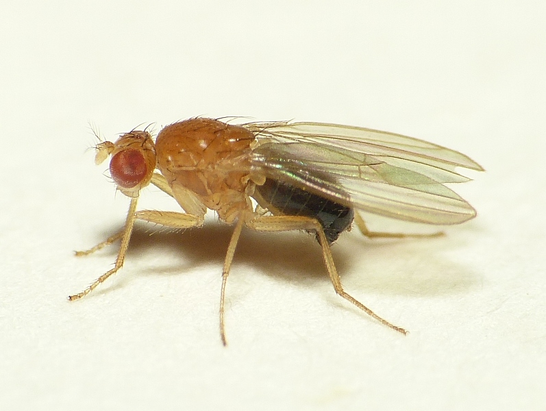
Despite their tiny brains, fruit flies have steep learning curves, as TU Delft researchers discovered using a technique that might one day shed light on neurological diseases.
A lot is happening in that tiny head. (Photo: Wikipedia)
Aversive therapy is best known for treating people with addictive behaviours, like those found in alcohol use disorder, but you can also use it to condition fruit flies. If you let these insects smell a sweet substance and then immediately give them an electric shock, they will remember that they should stay away from that odour.
You can find this out through behavioural experiments. But you can also peek inside the heads of these insects. The latter is what a team of researchers from TU Delft and the University of Oxford did. Their feat has pushed the boundaries of imaging.
In an article published in the scientific journal Elife, the researchers describe how they were able to pinpoint protein synthesis at subcellular resolution, right within the dendrites of neurons. Dendrites are the extensions of nerve cells that propagate the electrochemical stimulation received from other neural cells.
The researchers were able to localise mRNA molecules: the genetic material that encodes proteins. In this case, it was a protein called CaMKII, which plays an important role in a cascade of reactions that leads to the electrical firing of neurons.
‘Many neurological diseases are associated with proteins’
Animals and humans create memories by changing, strengthening, or weakening connections between neurons. How well they do so is defined by their so-called plasticity, their capacity for creating new connections. “Protein CaMKII plays a role in the plasticity,” says second author Carlas Smith, Assistant Professor at the Smart Optics Lab at the Delft Center for Systems and Control and Imaging Physics (AS and 3mE Faculties). “It is important that we get a better understanding of the role of proteins in the functioning of neurons. Many neurological diseases are associated with proteins. Chances are that similar proteins such as CaMKII also play a role in humans.”
As far as Smith knows, his team was the first to obtain images of single mRNA molecules within neurons in an intact brain at single-cell resolution. Smith was the principal investigator (research leader), yet would like to emphasise that most credit should go to Jessica Mitchell, a post doc in his team. “And you should also mention distinguished Oxford neuroscientist Scott Waddell, as one of the authors,” the TU Delft researcher adds.
Electric shocks
Right, so how did the researchers go about it? The imaging scientists put hundreds of fruit flies in transparent polyvinyl tubes with strong odours at one end. When in the vicinity of the scent, the insects received an electric shock through a small electrode. That experience set in motion a cascade of reactions in their brains, ultimately leading to memorisation of the event and aversive conditioning.
To find out how long it takes for the mRNA molecules, that play an important role in this process, to arrive at the scene (in the dendrites), the researchers extracted fly brains at different intervals after the olfactory learning experience. After this, they used a technique called single-molecule fluorescence in situ hybridisation, in which you use fluorescent dye to visualise molecules within single cells.
Smith is specialised in super-resolution microscopy. Ultimately he hopes to perform a similar trick in live fruit flies. He received a Veni grant from the Netherlands Organisation for Scientific Research a couple of years ago for super-resolution microscopy in live tissue.
Do you have a question or comment about this article?
tomas.vandijk@tudelft.nl


Comments are closed.