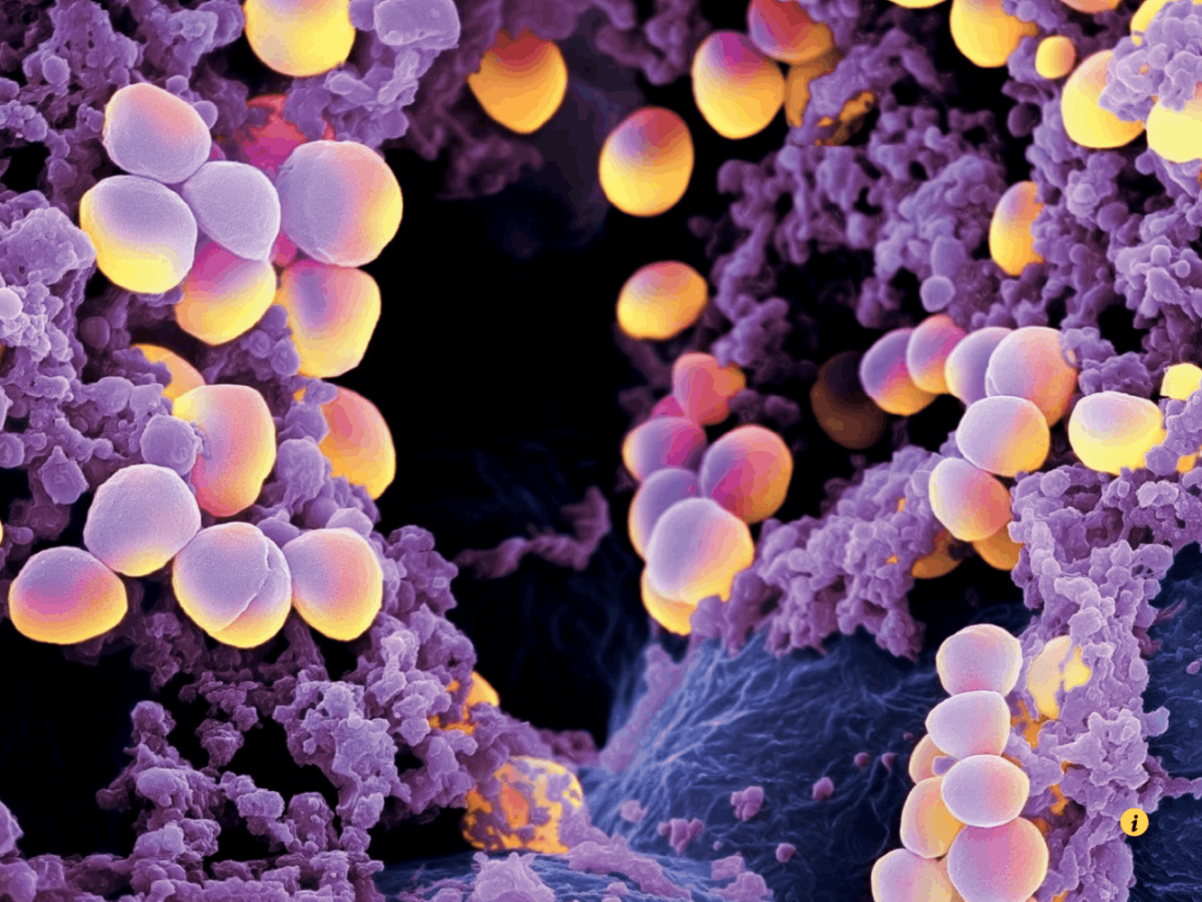
Historically, abscesses were believed to be identical and simply ‘big bags of pus’. But TU Delft researcher Raf Van de Plas and American colleagues showed that abscesses can have totally different molecular environments. Their work paves the way to new drug targets.
A team of researchers from the Vanderbilt University (US) and Dr Raf Van de Plas of the Delft Center for Systems and Control reveals how bacterial infection affects intact tissues. Their work delivers unprecedented insight into the spatial and molecular organization of bacterial infections, an aspect that can play a crucial role in the identification of new drug targets.
The scientists used a combination of imaging techniques to detect changes in the abundance and location of metal-binding proteins and nutrients in mouse tissue infected by Staphylococcus aureus, they reported in the March 14th edition of Science Translational Medicine.
“The study focused on a mouse model of dissemination of Staphylococcus aureus infection (or staph infection) to find the bacterial and host proteins that are found in and around abscesses, the area of inflammation around a bacterial infection”, says Raf Van de Plas, whose research focuses on the computational analysis of molecular and spectral imaging signals and systems.
‘This is an important finding’
Historically, abscesses were believed to be identical and simply “big bags of pus”, a Vanderbilt University press release states. The researchers however found that abscesses, even those in the same tissue, have different molecular environments. Some metals are excluded from all abscesses, and some metals are in one abscess, but not in another.
“This is an important finding”, says the Delft researcher. “Since metals are needed for essential biochemical reactions, the competition for metal between host and pathogen can be a determining factor in deciding the outcome of an infection. The body wages a “war” against the bacterial infection, resulting in distinct tissue zones in which different molecules play important roles.”
‘Developing new ways to combat bacterial infection is essential’
Being able to observe the strategies employed by the host attempting to thwart the infection, and the bacterial responses to these tactics, plays a crucial role in our understanding of bacterial infection dynamics. “In turn, this understanding is essential to developing new ways to combat bacterial infection” the Delft researcher adds. “This becomes increasingly important as the number of antibiotic-resistant bacterial forms grows and hospital-acquired infections form a growing health challenge.”
Over the past several years, the investigators have used various imaging modalities to study infection. Each approach has its own strengths. Magnetic resonance imaging (MRI), for example, shows how the physical anatomy changes in response to infection. Mass spectrometry reveals specific molecules with high sensitivity. Bioluminescence imaging (BLI) lets investigators study changes in gene expression in vivo.
“Using a single imaging technique to look at a tissue sample can provide very useful information, but such a singular viewpoint rarely delivers a comprehensive understanding on what’s going on. In multi-modal imaging studies, different imaging techniques have their own advantages and disadvantages, and deliver different complementary viewpoints on a phenomenon, connecting the dots.”
The work reported in this Science Translation Medicine paper fits into the broader cross-Atlantic collaboration that is being built out between Delft and the Vanderbilt university in Nashville, spanning across fields ranging from mathematical engineering and analytical chemistry to medicine and biology.
Do you have a question or comment about this article?
tomas.vandijk@tudelft.nl


Comments are closed.