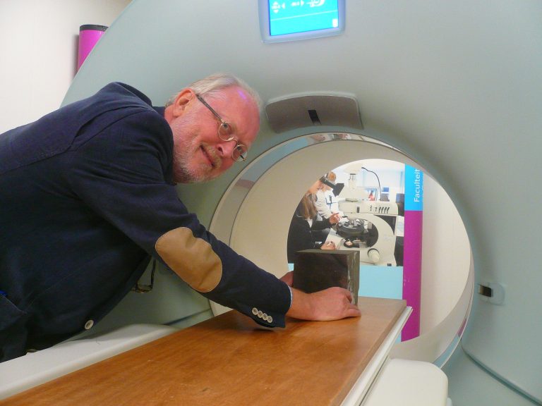
The new CT-scanner at the Faculty of Civil Engineering and Geosciences was made for humans. But it can dissect rocks just as easily.

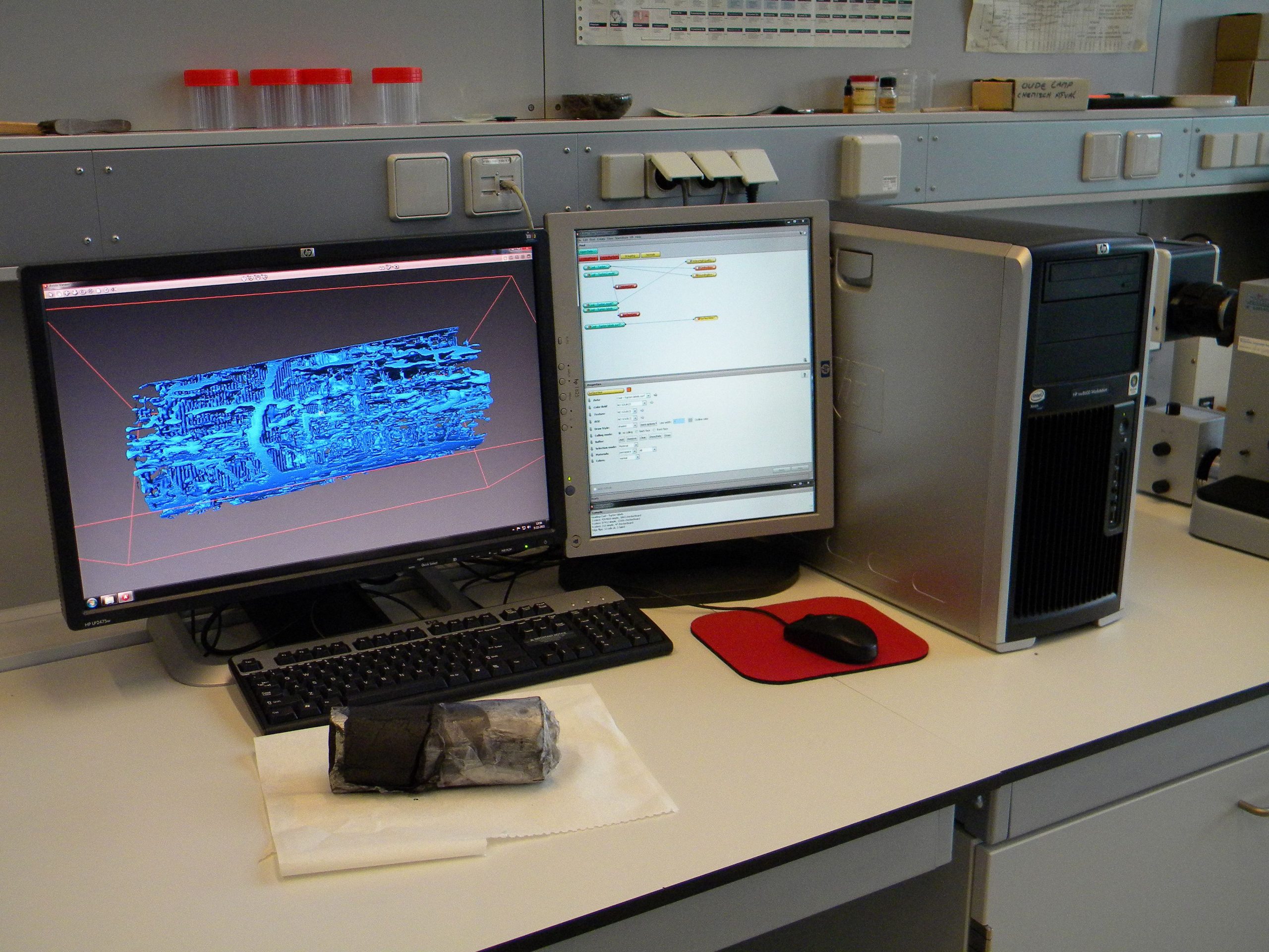
Lab technician Joost van Meel puts a composite cylinder on the scanner bed. He leaves the room and switches on the scanner. Inside the machine, and invisible to the eye, two heavy X-ray sources rotate around the sample, scanning every millimetre of it from every angle. Meanwhile, the bed translates slowly through the scanner hole. The new scanner makes 64 slides per millimetre while the resolution in the other directions is about 0.25 millimetres. Van Meel conjures up the image of a sandstone core in the cylinder. There is what seems to be a wet stain was entering the core from one side. Measuring the penetration speed of water through sandstone under pressures up to 300 bars is one example of the many applications that the scanner has.
“It can help you finding the best ways to get oil out of a certain rock”, says Dr Karl Heinz Wolf, associate professor at the Faculty of CEGS. If, for example, you want to produce oil from chalk, a very fine grained type of limestone, you may consider applying pressure or adding acid to detach the oil from the surrounding rock. The CT scanner shows what’s happening in the rock: wormhole type channels are formed. Do chemical reactions occur? Do we have different generations of wormholes? Such knowledge may be used to improve the geophysical models.

Other applications are gas winning from coal seams, extracting oil from tar sands or inspection of bricks (are they as solid as they look?). And that is all within the CEGS faculty. Others have shown interest in scanning a piece of wood of a million years old, or aluminium structures from Aerospace Engineering.
Wolf, who played an important role in acquiring the scanner for the university, said he was very glad about it. “It’s an asset for the experimental geoscience and engineering research”, he said. The role in education is limited to students reading the scans and trying to understand what information can be extracted from them.
Within CEGS, the CT-scanner nicely compliments the micro CT-scanner and Environmental Electron Microscope with a finer resolution but a sample size limited to a candy bar or less.
Researchers from other faculties are welcome to scan whatever samples they may have. The internal tariff varies from 100 (single shots) to thousands of Euros (entire campaign) to cover the operational costs of 40,000 Euros per year.
Do you have a question or comment about this article?
delta@tudelft.nl
Comments are closed.