This technique can make small creatures and biopsies transparent. The unique optics developed by PhD candidate Jelle van der Horst can look through 4 mm of tissue with infrared light.

The standard way of studying tissue samples from patients involves making cross-sections from the cylinder-shaped biopsy, staining them and putting the thin slices under the microscope to look for deviant and possibly malignant cells.
The new optic technology that Dr Van der Horst has developed at the Faculty of Applied Science has the potential to inspect an entire piece of tissue straight from the needle. It’s called OCPT or optical coherence projection tomography, and it uses highly advanced filtering techniques to keep out all the scattered light and use only one photon in a trillion (1012) that has travelled straight through the sample.
‘OCPT greatly increases imaging depth in tissue, improves contrast, and potentially allows for in vivo imaging’
The technology was tested on a piece of silicon rubber with a little white paint, and on a zebra fish fixed in agar. When a beam of light enters such an opaque object, most photons are scattered, randomly changing their direction of travel about every ¼ mm, while the sample is 4 mm in diameter. The denser the material, the shorter this so-called mean free path of photons will be. Still, it’s possible to detect this minute portion of the light going straight through the sample by comparing the light received with the light entering on the other side.
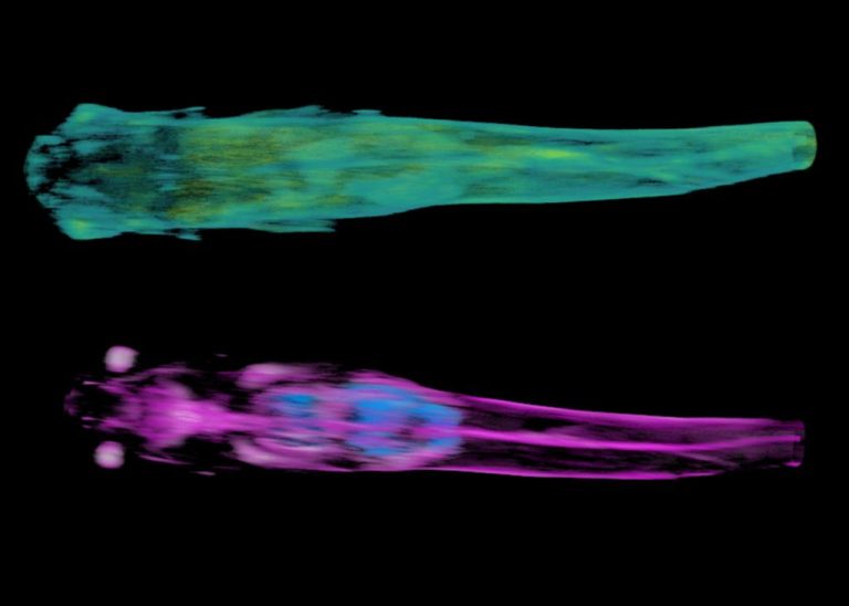
In fact, scanning line by line, two different images can be reconstructed. These images are based on the total weakening of the light, called attenuation, and on the refractive index of the material. The images seem to contain different information about the material properties of the tissue. The refractive index, for example, appears to be very sensitive to the air in the swimming bladder (blue in the second image).
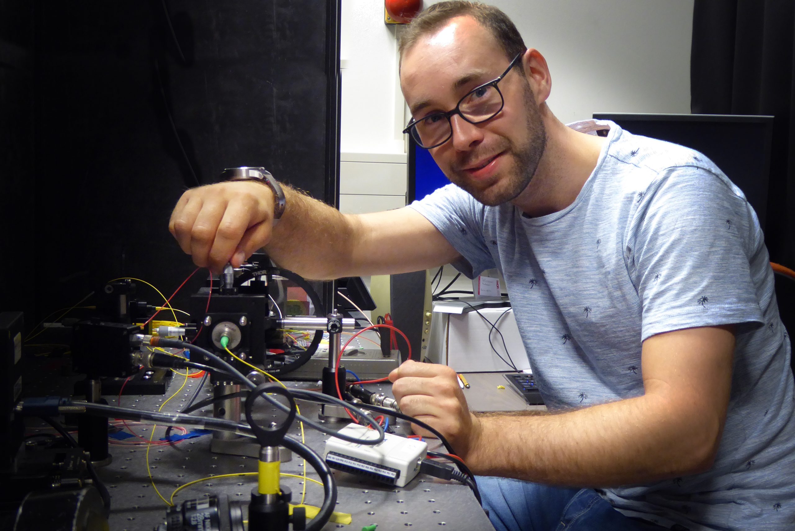 Jelle van der Horst (Photo: Jos Wassink)
Although the 3D measurements currently take several hours of imaging, the process can be speeded up by deploying rapid beam-scanning techniques and by reducing the number of angles for each image (currently 180 images are made with 1 degree of rotation between takes).
With a micrometre resolution for several millimetre-sized objects, OCPT fills a gap between other 3D-imaging techniques such as optical coherence tomography (up to one millimetre-sized objects) and photoacoustic tomography.
- Jelle van der Horst, High-resolution deep-tissue quantitative optical tomography, PhD supervisors Professor Lucas van Vliet and Dr Jeroen Kalkman (Applied Sciences).
Do you have a question or comment about this article?
j.w.wassink@tudelft.nl
Comments are closed.