The G-SPECT scanner, developed by MILabs, received the Commercial Innovation of the Year award at the 2015 World Molecular Imaging Congress on Hawaii on September 17, 2015.

MILabs’ founder Professor Freek Beekman heads the section Radiation Detection and Medical Imaging at the Faculty of Applied Sciences.
SPECT stands for Single Positron Emission Computer Tomography. It is a radiological 3D-imaging method that works with radioactive isotopes that bind to specific organs and tissues, such as tumors. SPECT allows oncologists to obtain 3D-images of cancers for diagnosis or treatment. Known drawbacks of SPECT are the fuzzy images (lack of resolution) and the long scanning times.
G-SPECT, developed by the Utrecht University spin-off MILabs, promises to improve on these drawbacks. The resolution is less than half of the conventional machines (2.5 instead of 7 millimetres) and the scanning times are less than a minute for most cases, compared to 10-30 minutes for the current machines.
The improvement is due to the full-ring detector instead of having a set of smaller detectors rotating the patient to do the sampling. The ring allows for a simpler construction simpler and a quiet operation. And because of the higher sensitivity, the acquisition times are so much lower (in the order of a few seconds) that dynamic imaging becomes possible to see for changes in the blood flow for example.
“It’s not that we always get that one-second time resolution, but in some cases it is possible,” said Beekman in an article on Medical Physics Web . “It’s hard to say what the scan speed will be in patients, but it will be much faster than you can achieve with a normal clinical system where the detector has to rotate around the patient.”
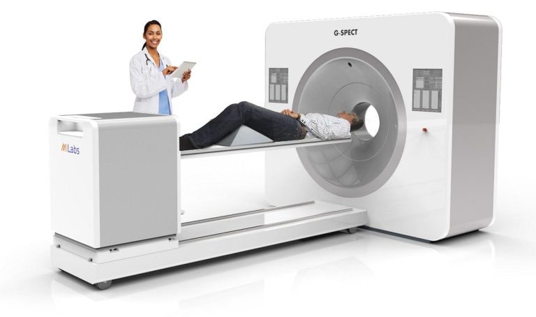
The current scanner only has a small opening, allowing only scans of brain or limbs. MILabs is working on another scanner with a larger opening that will enable total body imaging and high-resolution functional heart imaging.
Beekman suggests that this win at the WMIS conference may in part be due to frustration with existing technology and a real clinical need for improvement. “Today, millions of people get SPECT scans, and all those scans have limited resolution, which lends uncertainty to diagnosis in many cases,” he explained. “All the infrastructure is there, nuclear medicine departments have rooms ready for SPECT, but there’s a huge problem in that they cannot see sharply with their existing SPECT systems. We’ve had a large interest in the new scanner.”
He emphasized that G-SPECT is still awaiting approval for clinical use and currently, can only be sold for preclinical large animal applications. “We are working hard on this process and hope to get approval to sell systems next year – early next year I hope,” said Beekman.
Do you have a question or comment about this article?
delta@tudelft.nl
Comments are closed.