Flow experts have done calculations on the blood vessels in the brain. This provides information that an MRI scan cannot provide such as vulnerable areas in vessel walls.

During a MRI brain scan the head is immobilised. (Photo: National Cancer Institute via Unsplash)
The group of Prof. Saša Kenjereš (Faculty of Applied Sciences, AS) enjoys working on flows in biomedical environments such as air through the lungs and blood through the heart. Recently, Kenjereš and his PhD student Romana Perinajová published a study on the complex flows in a roundabout of arteries below the brain that supply blood to the brain.
“The problem is that you often don’t know whether your simulation is correct,” Kenjereš explains. “Many people calculate simplified systems flows through channels or tubes. That is standard. Our challenge is to calculate the flow in a complex system of blood vessels with branches and merges. In collaboration with the Amsterdam UMC, we were able to compare our flow calculations with MRI images.”
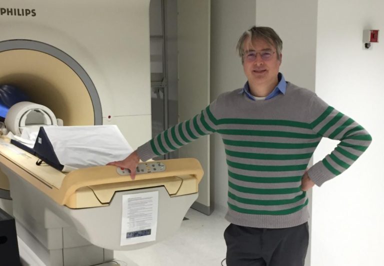
Pim van Ooij (Amsterdam UMC) at the MRI-scanner used for the publication. (Photo: private collection)
Clinical Engineer Pim van Ooij of the Amsterdam UMC says “I work with 4D flow MRI (successive 3D images, JW). This allows us to map blood flows in the brain very well. Another technique that we can use is CFD – computational fluid dynamics. CFD is a numerical simulation with boundary conditions and assumptions, while MRI is a measurement. But we cannot measure as well as in CFD simulations.”
Stresses and complaints
As part of the collaboration, Van Ooij provided blood vessel geometry (from a 25-year-old volunteer) and average flow velocities. Perinajová and Kenjereš used CFD to calculate the flow in detail, especially near the vessel wall. Kenjereš explains that “It all depends on the thickness of the boundary layer, but close to the wall the voxels (three dimensional cubes the computer uses for its calculations, JW) are very small, something like a hundredth of a millimetre. This allows us to calculate the forces on the vessel wall very precisely.”
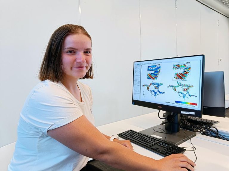
PhD student Romana Perinajova performed the CFD computations. (Photo: private collection)
This is of interest to vascular specialists because vessel wall strain is linked to health problems. In places where the force on the vessel wall (shear stress) is low, blood flows slowly and deposits can easily form (atherosclerosis). Places with high shear stress, and thus high flow velocity along the vessel wall, are prone to damage and sometimes bulges (aneurysm).
Flow analyses clearly show these spots (see video.)
Calculated flow velocities at the junction of cerebral arteries (Circle of Willis). The camera first zooms in on the blue area with low shear stress (risk of plaque formation) and then on a red area with high stress vessel wall (risk of aneurysm). Video: Romana Perinajová, Kenjereš Lab, TU Delft.
By combining flow analysis with biochemistry, local oxygen concentrations can even be calculated. One application is identifying sections of blood vessels that have lacked oxygen for long periods. These spots are shown in red in the video below.
The combination of MRI and flow analysis (CFD) provides the basis for calculating oxygen transport and identifying spots of oxygen deficiency. Video: Romana Perinajová, Kenjereš Lab, TU Delft.
Smart scanner
Now that CFD appears to add relevant information to MRI images, the question is how to proceed given that detailed 3D flow calculations are notoriously demanding in terms of computing power. Kenjereš’ estimate is 10 million voxels with six variables, thousand time steps per heartbeat and 3-5 heartbeats per analysis. That’s a few days of computation, or a few hours on a supercomputer.
The researchers are now turning their hopes to artificial intelligence. A suitable neural network trained with a few dozen examples could, they say, predict flow rates very well at a fraction of the computing time. This would make the flow analyses by Kenjereš and his group clinically applicable as an extension of the MRI scan.
“In the future, you won’t need to do CFD at all,” Kenjereš says , “if we make good time series, they can be used to train AI systems that automatically recognise hotspots and identify spots with increased risk of pathologies.”
- Read the publication at the Royal Society: On the identification of hypoxic regions in subject-specific cerebral vasculature by combined CFD/MRI
Do you have a question or comment about this article?
j.w.wassink@tudelft.nl
Comments are closed.