Diabetes patients need to have their eyes checked regularly to monitor the progression of retinal degeneration. Dr Kedir Adal developed a computer-aided diagnosis tool.

Kedir Adal op postersessie. (Foto: Privécollectie Adal)

In the Netherlands alone, there are almost one million people with diabetes. One of the most common complications of this metabolic disorder is that it affects blood vessels in the retina. Called diabetic retinopathy (DR), small lesions from blood vessels in the retina can result in loss of vision or even blindness. To monitor the onset and progression of DR, people with diabetes need their eyes to be regularly checked by trained experts.
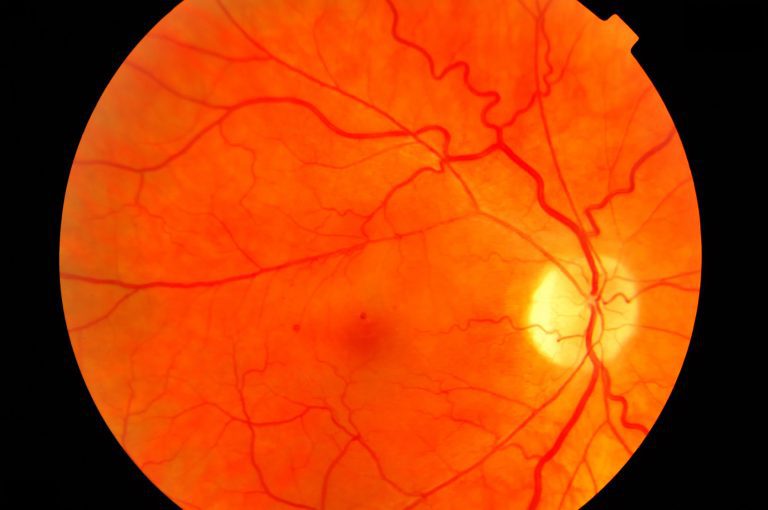 Fundus photo. (Photo: Eye Hospital Rotterdam)
This is done by taking retina or ‘fundus’ photos and depending on the analysis of the photos, the expert will either schedule a follow-up examination or refer the patient to an ophthalmologist (eye specialist).
The screening is time consuming, subjective, and does not use images from previous screenings to monitor disease progression, says Dr Kedir M. Adal. Finding relevant abnormalities in a photo of the retina is so taxing that the previous year’s photos for comparison are rarely dug up. How could a comparison even be done? The illumination is different and the field of vision is not the same. And yet, changes in the retina over time contain valuable information on the development of DR.
By processing the fundus images with computer algorithms, data scientist Dr Kedir M. Adal has removed these obstacles. He developed an automated DR diagnosis tool during his PhD research at the TU Delft Faculty of Applied Sciences, in collaboration with the Rotterdam Ophthalmic Institute (ROI), the Rotterdam Eye Hospital’s research institute. His computer aided diagnosis tool paves the way for monitoring the development of diabetic retinopathy over time. In his talk before his PhD defence he outlined the steps he had taken.
- Normalisation of the illumination (converting the orange image into a high contrast image with grey tones).
- The Rotterdam Eye Hospital’s protocol is to take four images (top, bottom, left, right) for each eye to show the maximum surface of the retina. Image processing merges the four images into a single mosaic by zooming, rotating, translating, and assembling the images onto a fixed register.
- Comparing the new processed image with previous images such as those from the year before, and highlighting the differences.
- Determining the clinical relevance of the changes with a trained neural network.
1: fundus mosaic, 2. white spots mark differences, 3. red marks on clinical relevant changes. (Gif: Kedir Adal)
No additional hardware is required to run his Computer Aided Assessment, Adal explained. His system is software-based and runs on a laptop.
Dr Koen Vermeer, Adal’s PhD supervisor from the Rotterdam Ophthalmic Institute, sees clear applications for the new algorithms. ‘They can generate a risk profile for the patient which can be used for behavioural changes, or for adjusting the time until the next fundus photos. Low risk, long time. High risk, short time,’ Vermeer wrote in an email. ‘Another application could be educating patients by showing them changes in the retina. This information may motivate patients to change their lifestyles.’
Will the system be introduced in the screening programme at the Rotterdam Eye Hospital? Well, that is easier said than done. The Rotterdam Eye Hospital no longer does screening. Furthermore, because health care is financed on the basis of cure and not prevention, there is no budget for developing preventive technologies such as Dr Adal’s algorithms.
Meanwhile, a huge research programme on DR has been launched in the US. Motivated by the sheer number of people involved, and the role of deep learning in interpreting fundus images, large hospitals and IT giants such as Google have embarked on a large scale research programme.
‘For a relatively small group such as ours, it has become difficult to compete for this application,’ writes Vermeer.
- Kedir M. Adal, Computer-Aided Assessment of Longitudinal Fundus Photos for Screening Diabetic Retinopathy, PhD Supervisors Prof. Lucas van Vliet (Applied Sciences) and Dr Koen Vermeer (ROI), 12 March 2018.
A short video shows the process (video by Kedir Adal)
 Kedir M. Adal. (Photo: Hans Krüse)
Data scientist Kedir M. Adal was born in Ethiopia in 1985. He did his BSc in Electrical Engineering in 2006 at the Mekelle University in Ethiopia, followed by a MSc in computer vision and robotics in Bourgogne (France, 2012). In February 2013 he started his PhD project with TU Delft, the Rotterdam Eye Hospital and the Rotterdam Ophthalmic Institute. He now works at ASML in Eindhoven.
Do you have a question or comment about this article?
j.w.wassink@tudelft.nl
Comments are closed.