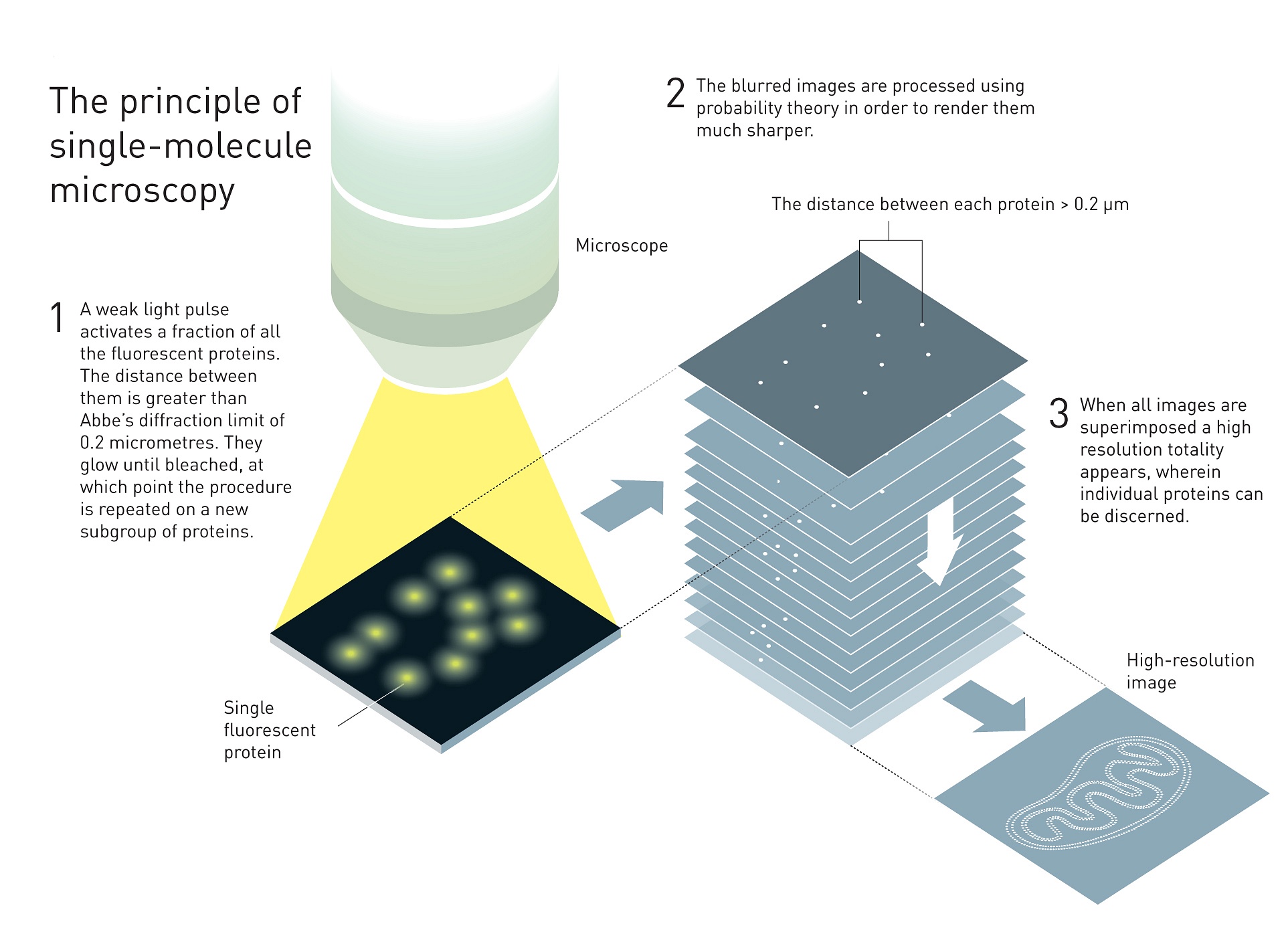
A combination of techniques which has enabled researchers to image single molecules with optical microscopes, called ‘nanoscopy’ or ‘superresolution microscopy’, has been awarded the Nobel Prize 2014 for chemistry.
‘For a long time optical microscopy was held back by a presumed limitation: that it would never obtain a better resolution than half the wavelength of light’ says the Nobel Prize committee in their press release. ‘Helped by fluorescent molecules the Nobel Laureates in Chemistry 2014 ingeniously circumvented this limitation. Their ground-breaking work has brought optical microscopy into the nanodimension.’
The winners are Eric Betzig, Stefan Hell and William Moerner who each developed a part of the intricate optical technique.
Stefan Hell (Germany) was obsessed by the fact that optical microscopy couldn’t produce details smaller than about 250 nanometer (0,2 micron) – half the wavelength of the light used – called the Abbe-limit. He figured that he could use single fluorescent molecules as tiny nano-sized light sources. In order to make only single molecules light up instead of the whole batch, he converted a laser into a nano-flashlight with which he scanned a sample line after line. This technique was called stimulated emission depletion or STED and it was developed at the university of Turku, Finland. In 2000 he showed that the technique actually worked by imaging an E.coli bacterium shaper as ever before. A Nature news feature from 2009 spoke of ‘microscopic marvels: the glorious resolution’
Hell couldn’t have developed his STED microscopy if it wasn’t for the possibility of measuring the light emission by a single molecule – an achievement that W.E. Moerner set in 1989 when he was working at the IBM research centre in San Jose, California. Later, in 1997, he published his findings of a special variant of the much used green fluorescent protein (GFP) which could be turned on and off at will. Light with wavelength of 488 makes the protein glow once and fade away for good. However, it could be revived with slightly bluer light. In a Nature publication, he showed that individual molecules could be imaged like tiny lamps.
Erik Betzig combined the switchable proteins and the STED microscopy into the superresolution technique in use today. It uses weak light so that only a small number of fluorescent markers in the sample lights up (and subsequently fades to black). The optical microscope captures a number of tiny lights and stores the image. The procedure is repeated over and over again until the superimposed tiny light dots (far smaller than Abbe’s limit) form a high-resolution image of the cell or organelle that was labelled.
The three Laureates are still active as researchers. Stefan Hell has peered inside living nerve cells in order to better understand brain synapses. W. E. Moerner has studied proteins in relation to Huntington’s disease. Eric Betzig has tracked cell division inside embryos.
‘The Nobel Laureates in Chemistry 2014 have laid the foundation for the development of knowledge of the greatest importance to mankind’, says the committee.
In Delft, superresolution microscopy is applied by Dr. Bend Rieger and Dr. Sjoerd Stallinga who recently published on a technique for measuring distances in optical nanoscopy.


Comments are closed.